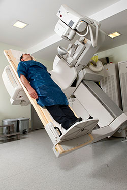
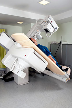
Fluoroscopy is an imaging technique that uses a steady beam of X-rays, occasionally with the use of contrast dye, to observe the real-time movement of an organ or body system. Using fluoroscopy, doctors and radiologists can view many systems of the body, including:
- Skeletal systems
- Digestive systems
- Urinary systems
- Reproductive systems
At several of our Imaging Center locations across the Greater Houston area, Memorial Hermann offers a full range of professional X-ray exams, including fluoroscopy exams, that are performed by specialty-trained radiologists.
If your doctor has ordered a fluoroscopic imaging exam, contact us today. We can answer any questions you may have about the procedure, verify your insurance coverage and schedule your appointment at a time and location that’s convenient for you.
When is Fluoroscopy Commonly Prescribed?
Fluoroscopy can be used alone or in conjunction with other imaging modalities to perform a wide variety of diagnostic examinations and procedures. A few common uses include:
- The placement of devices into the body, such as stents, which are used to open narrow or blocked blood vessels and arteries
- Angiograms — a diagnostic examination that uses a fluoroscope and contrast dye to visualize blood vessels and organs
- Conducting orthopedic surgery using real-time X-ray images to guide joint replacements and treat fractures
- Visualizing the upper and lower gastrointestinal tract to determine the cause of intestinal-related problems, such as acid reflux, difficulty swallowing or abdominal pain
How Does Fluoroscopy Work?
Fluoroscopy is similar to an ultrasound, in that it produces real-time images of structures inside the human body. However, unlike ultrasound – which uses high-frequency sound waves to produce images – fluoroscopy uses the same type of ionizing radiation found in traditional X-ray imaging.
The name “fluoroscopy” comes from fluorescence, which pertains to the use of certain substances to convert absorbed electromagnetic radiation into visible light. In many cases, this refers to the use of contrast agents which can be administered orally, intravenously or through an enema. These substances appear opaque during imaging and can help highlight certain organs or structures that may be difficult to capture using traditional X-ray imaging, such as blood vessels, arteries and the gastrointestinal (GI) tract.
Types of Fluoroscopy Exams
Fluoroscopy is a functional form of X-ray imaging that can be used in a variety of ways. The following are just a few examples of diagnostic application of fluoroscopy.
- Barium X-rays use a barium-based contrast material to examine the upper and lower gastrointestinal (GI) tract. This procedure helps detect abnormalities of the intestines and is often ordered to investigate symptoms such as pain, constipation or blood in the stool.
- Arthrography is used to help diagnose conditions of the joints. This procedure is effective for detecting disease within ligaments, tendons and cartilage. Indirect arthrography involves contrast material injected into the bloodstream, whereas direct arthrography uses injections into the joint itself.
- Hysterosalpingography examines the uterus and fallopian tubes of women who are having difficulty becoming pregnant. It can also be used to investigate abnormalities within the uterus, and determine the presence and severity of masses, adhesions and uterine fibroids. In some cases, this procedure can actually open blocked fallopian tubes and aid the patient in becoming pregnant.
Other forms of fluoroscopic imagery, such as cardiac catheterization, vertebroplasty and kyphoplasty, are not performed in outpatient imaging centers. These are routinely utilized in interventional radiology (IR) labs or surgical suites. Cardiac catheterization and vertebroplasty/kyphoplasty are not performed in our outpatient imaging centers. They are routinely performed in IR labs or surgical suites.
Benefits and Risks of Fluoroscopy
Like most X-ray procedures, fluoroscopy carries some risks. Because radiation doses received depend on the imaging study performed, it’s possible to be exposed to higher doses of radiation through complex interventional procedures that require extended periods of fluoroscopy. It is important to note that each patient’s individual risk for side effects from fluoroscopy is statistically very low.
According to the FDA, the following radiation-related risks associated with fluoroscopy include:
- Injuries to the skin and underlying tissues, which may occur shortly after exposure
- Radiation-induced cancers, which may begin to develop months or years after exposure
The clinical benefits of X-ray imaging exams deemed medically appropriate (i.e., potentially lifesaving) far outweigh the small risk of radiation exposure. In order to follow the standards for care set forth by the FDA's Initiative to Reduce Unnecessary Radiation Exposure from Medical Imaging, fluoroscopic exams must be performed with the lowest acceptable radiation levels for the shortest time necessary. The performing radiologist will meticulously document the total amount of radiation you receive in order to help minimize your overall lifetime exposure levels.
If you have any questions or concerns about which imaging technique is right for you, contact a Memorial Hermann Imaging Center staff member today. We will be happy to assist you.
What to Expect from a Fluoroscopy Exam
The complexity of your fluoroscopic imaging exam will vary depending on the underlying reason for the examination. For example, some fluoroscopic exams are simple outpatient procedures, such as imaging the upper gastrointestinal tract or conducting a barium enema to examine the colon. However, other fluoroscopic exams may involve the use of general anesthesia or sedation, such as cardiac catheterization to examine the heart and coronary arteries, or as part of orthopedic surgery to align and set fractured bones.
If your doctor has ordered a fluoroscopy exam, you may be wondering what to expect. The following information will provide some background on how you should prepare for your imaging study, what to expect during the exam and how long it will take to receive your results.
Preparation
Perhaps the best way to prepare for a fluoroscopic imaging exam is to first talk with your doctor. According to FDA guidelines, patients and parents of children undergoing X-ray imaging exams such as fluoroscopy should do the following:
- Keep track of medical imaging histories with your referring physician, and make sure to double-check them when a new exam is recommended
- Inform your doctor if you are pregnant or think you may be pregnant
You should also talk with your referring physician about the benefits and risks of recommended imaging procedures such as fluoroscopy. The following are some sample questions to help start this discussion:
- How will the results of the exam be used to evaluate my condition or guide my treatment (or that of my child)?
- Are there alternative options for imaging that do not use ionizing radiation?
- Does the imaging facility use techniques to reduce radiation exposure, especially to sensitive populations such as children?
The Day of the Fluoroscopy Exam
Fluoroscopic exams vary depending on the type of imaging exam requested by your referring physician. Some exams may require you to fast for several hours ahead of time, while others may not, and some may require an IV of contrast dye, or a barium enema. In order to adequately prepare for your fluoroscopy exam, talk with your doctor ahead of time.
When you first enter a Memorial Hermann Imaging Center, you will need to check in at the front desk before completing routine billing paperwork in the business office. An imaging technologist will then come to collect you from the waiting room and bring you to a private exam room. There, the technologist will review your information with you, explain how the exam will work and answer any of your questions.
Jewelry, piercings and other metal objects will need to be removed if possible, and you may be asked to wear loose clothing or change into a hospital gown. The imaging technologist will have you lie on the examination table for the duration of the exam and may ask you to change positions in order to get a clear image.
How Long Does a Fluoroscopy Exam Take?
At Memorial Hermann, the experienced staff members understand that your time is important, and we do our best to make sure each exam is quick and thorough. Please know that certain fluoroscopic exams may take longer than others – anywhere from 15 to 20 minutes, up to several hours for complex procedures (or if you are given a contrast solution that must be partially digested before scanning).
When Can I Expect My Results?
Images from your fluoroscopic imaging exam will be sent for immediate analysis by a professional radiologist. Once the radiologist has interpreted the images, a full report will be sent to your referring physician. This process usually takes about 2-3 business days. Your doctor may schedule a follow-up appointment with you to go over the results.
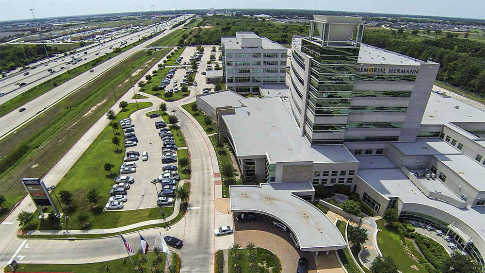
Choose Memorial Hermann for Convenient, Professional Fluoroscopy in Houston
Memorial Hermann Imaging Centers offer fluoroscopy at over a dozen locations throughout the Greater Houston area:
-
Memorial Hermann Imaging Center at Katy HospitalGet Directions23920 Katy Fwy Ste 120, Katy, TX 77494
-
Memorial Hermann Imaging Center at Rehabilitation Hospital - KatyGet Directions21720 Kingsland Blvd Ste 102, Katy, TX 77450
-
Memorial Hermann Imaging Center at Memorial City Medical CenterGet Directions925 Gessner Rd Ste 200, Houston, TX 77024
-
Memorial Hermann Imaging Center at Northeast HospitalGet Directions18955 N Memorial Dr Ste 100, Humble, TX 77338
-
Memorial Hermann Imaging Center - PearlandGet Directions10905 Memorial Hermann Dr Ste 104, Pearland, TX 77584
-
Memorial Hermann Imaging Center at Southwest HospitalGet Directions7789 Southwest Fwy Ste 150, Houston, TX 77074
-
Outpatient Imaging at Memorial Hermann | Rockets Orthopedic HospitalGet Directions5410 West Loop South, Bellaire, TX 77401

A physician order is required for a fluoroscopy scan.
Need to find a physician? Click here.
Schedule a fluoroscopy scan by calling (877) 704-8700.